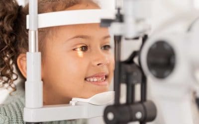
Are you worried about why your child’s vision keeps getting worse each year? Do you want a clear explanation of what’s happening inside their eyes, not just a stronger glasses prescription? At Beyond Eyecare, we use the latest imaging tools to track and manage myopia with precision.
I’m David Truong, Principal Optometrist at Beyond Eyecare in Sydney. In this article, you’ll learn all about how myopia is measured, the difference between axial and refractive myopia, and why tools like OCT scans give us an early look at eye health risks.
For parents wanting to stay ahead of their child’s eye health, here’s what to know and what to do next.
Key Takeaways
- OCT scans let us catch internal eye changes early.
- Axial length measurement is a more reliable progression marker than prescription alone.
- Refractive and axial myopia require different monitoring approaches.
- Devices like the REVO FC combine OCT with axial tracking for precise care.
Myopia And Its Impact On Eye Health
Myopia, also known as short-sightedness, occurs when the eyeball grows too long from front to back. As a result, light entering the eye focuses in front of the retina instead of directly on it. This causes distant objects to appear blurry while near vision remains clear.
This condition typically starts in childhood, especially during the early school years, and tends to worsen until the eye fully matures in early adulthood. Children with one or both parents who are myopic are more likely to develop the condition themselves.
Types Of Myopia
| Type | Description | Health Risks |
| Refractive Myopia | Steep corneal curvature or excessive lens power | Generally lower risk |
| Axial Myopia | Elongation of the eyeball | Higher risk of retinal detachment, macular degeneration, and other structural eye issues |
There are two main categories of myopia: refractive and axial.
Refractive myopia
This is caused by excessive focusing power in the eye’s front structures, typically a steep cornea or strong natural lens. This bends light too sharply, leading it to focus in front of the retina. Refractive myopia can often be corrected with glasses or contact lenses and typically carries a lower structural risk.
Axial myopia
This is when the eyeball elongates beyond its normal shape. This type is more common in children with worsening prescriptions and tends to develop and progress through school-age years.
Axial elongation stretches the internal structures of the eye, increasing the chance of retinal thinning, detachment, or other degenerative conditions later in life. Even if a child’s glasses stay the same for a year, the eye may still be growing.
That’s why axial length tracking is so important; it picks up changes that basic refraction misses.
Refractive Myopia vs Keratoconus
Optical Coherence Tomography (OCT) is a valuable tool in detecting and monitoring myopia, especially when distinguishing between axial and refractive causes.
In refractive myopia, OCT can help rule out structural abnormalities by confirming normal retinal and optic nerve anatomy.
It’s also useful in identifying subtle signs of early keratoconus, a condition that can mimic or contribute to myopic blur. Although keratoconus is primarily diagnosed with corneal topography, OCT pachymetry maps can reveal localized corneal thinning patterns.
Detecting keratoconus early is essential, as standard myopia treatments — like glasses or soft contacts — may be inappropriate for these patients.
| Feature | Refractive Myopia (Non-Axial) | Keratoconus |
|---|---|---|
| Cause | Excessive optical power due to a steep cornea or powerful lens, with normal axial length | Progressive corneal thinning and conical protrusion leading to irregular astigmatism |
| Prevalence in Australia | Specific data on refractive myopia prevalence in Australia is limited; however, overall myopia prevalence is increasing | A study from the Raine Study in Western Australia reported a keratoconus prevalence of 1.2% among 20-year-olds, indicating a higher prevalence than previously thought. Source |
| Progression | Typically stable; may increase with age due to lens changes | Often progressive, especially in adolescents and young adults |
| Astigmatism Type | Regular and symmetrical | Irregular and asymmetric |
| Detection Methods | Standard refraction; corneal topography or tomography if indicated | Requires corneal topography or tomography for accurate diagnosis |
| Management | Glasses, contact lenses, or refractive surgery | Specialty contact lenses, corneal cross-linking, or surgical interventions in advanced cases |
| Associated Risks | Generally low; primarily visual discomfort | High risk of visual impairment if untreated; potential for corneal scarring and vision loss |
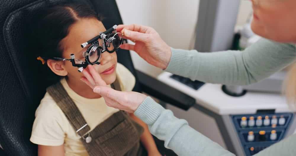
How Is Myopia Measured?
Myopia usually worsens over time. As a child’s eyeball continues to stretch, it increases the risk of complications later in life, including retinal detachment, macular degeneration, glaucoma, and myopic maculopathy. These risks grow exponentially once axial length exceeds certain thresholds, typically around 26mm.
That’s why early and precise measurement is critical, even when a child’s vision seems otherwise fine with glasses.
The Role Of Axial Length In Myopia Monitoring
Axial length is the distance from the front surface of the cornea to the retina at the back of the eye. Unlike changes in glasses prescriptions, which can fluctuate slightly due to measurement conditions, axial length provides a stable and objective metric of actual eye growth.
Tracking this measurement over time allows us to detect whether a child’s myopia is truly stabilising or still progressing, often before changes are visible in a standard eye chart test.

Using OCT Scans To Assess Myopia
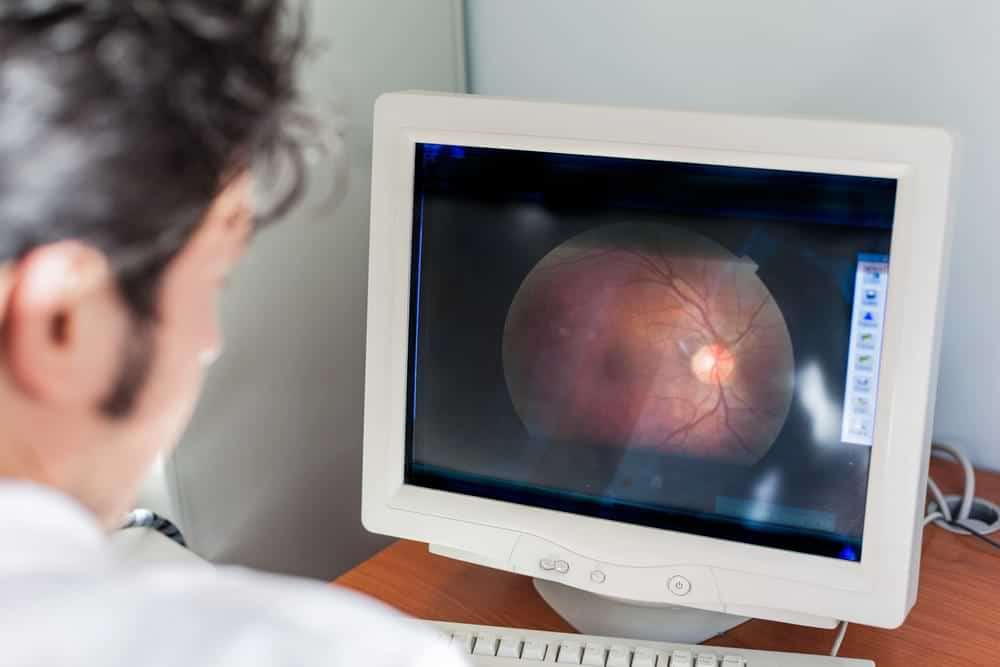
OCT, or Optical Coherence Tomography, is a non-invasive imaging test that uses light waves to capture high-resolution cross-sections of the retina. It measures the thickness of each retinal layer, the contour of the fovea (the central focus point of the retina), and the condition of the optic nerve head.
In the context of myopia, OCT scans are essential for identifying early signs of damage, such as myopic maculopathy, choroidal neovascularisation (CNV), or posterior staphyloma. These changes can appear silently (without symptoms) especially in cases of high or rapidly progressing myopia.
How OCT Scans Help In High Myopia
High axial myopia stretches the retina, making it more vulnerable to degeneration. Even in young patients, subtle signs of stress on the retina can be detected through OCT before they cause noticeable vision issues.
OCT imaging allows us to compare scans over time, track retinal thinning, and monitor for microstructural changes. This gives us a clearer picture of whether a treatment plan is working or needs to be adjusted. It also helps determine when to refer a child for specialist care, particularly if there’s concern about CNV or other sight-threatening complications.
Reading An OCT Scan
Interpreting an OCT scan involves assessing colour-coded maps of reflectivity. Red and orange areas typically indicate high reflectivity, such as dense tissue like the retinal pigment epithelium (RPE), while green and blue areas show lower reflectivity.
We use these visual cues alongside numerical data and previous scan comparisons to assess progression or response to treatment. A healthy retina has well-defined layers and stable measurements over time.
How myopia is measured will reveal various signals such as like posterior bowing, vitreoretinal traction, or peripapillary atrophy, features that indicate a higher risk for long-term visual complications.
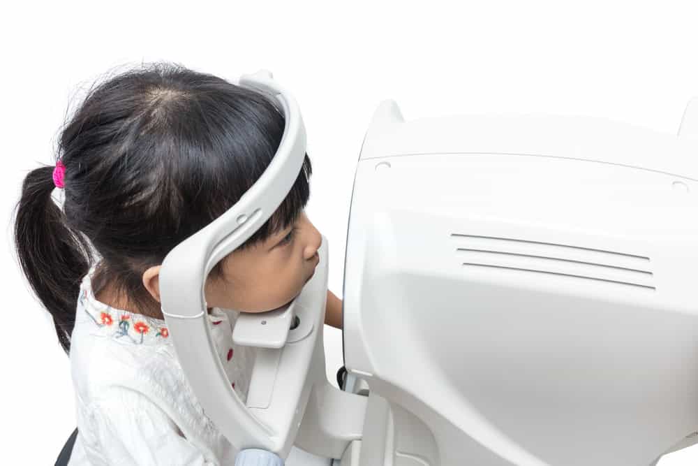
Tools Used To Measure Axial Length
At Beyond Eyecare, we’re committed to delivering the most precise care possible—which is why we’ve invested in the REVO FC, a cutting-edge diagnostic device that combines high-resolution OCT with a non-mydriatic fundus camera. This powerful tool plays a vital role in myopia management by allowing us to track axial length changes, assess retinal and corneal structure, and detect early signs of complications like keratoconus or myopic maculopathy.
What makes the REVO FC especially helpful for children is its AccuTrack™ real-time eye tracking system. It compensates for blinks and eye movements, making it easier to scan young patients who struggle to sit still. With fully automated alignment, focus, and capture, we can obtain accurate, high-quality images with minimal patient cooperation. This technology helps us monitor your child’s eye health more effectively, ensuring timely and personalised myopia control strategies that grow with them. It’s all part of our commitment to smarter, more comfortable care.
What Parents Should Know About How Myopia Is Measured
Many parents assume that if their child’s prescription isn’t changing, the eye must be stable. But that’s not always true. Prescription can plateau while the eyeball continues to grow. Axial length reveals this hidden progression. That’s why we encourage all families considering myopia control to request axial tracking as part of their child’s eye exams.
Questions To Ask Your Eye Doctor
Don’t hesitate to ask about the technology being used to monitor your child’s eyes. Key questions include:
- Do you measure axial length at each visit?
- How often do you perform OCT scans?
- What changes in axial length would prompt a shift in the treatment plan?
Having these conversations ensures you’re getting the most informed care. At Beyond Eyecare, we believe that thorough, ongoing monitoring leads to better outcomes, and more peace of mind for parents.
Final Thoughts
Myopia is beyond having blurred vision. It’s also a structural issue with lifelong risks. That’s why we use tools like OCT imaging and axial length measurement to guide care with accuracy. The sooner we start tracking eye changes properly, the better we can protect long-term vision.
At Beyond Eyecare, we offer modern diagnostics, evidence-based strategies, and clear guidance on how myopia is measured for families who want the best for their children’s long-term eyesight.
Book an appointment with Beyond Eyecare today at Zetland (02) 9662 6364 or Surry Hills (02) 9556 1160. You can also schedule a convenient time through our website.

Therapeutical endorsement